Temporal Tendinitis
 Temporal tendonitis (or, tendinitis) is perhaps one of the most common craniofacial pain disorders seen in clinical practice. Unfortunately, this widespread problem is frequently confused and misdiagnosed as an intra-articular (within) the temporomandibular joint disorder.
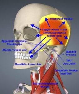
The “temporal tendon” connects the jaw bone to a muscle that spans the side of the head (the “temporal muscle”). The purpose of the temporal muscle is to pull the temporal tendon, which helps the mouth to close.
The condition “temporal tendinitis” refers to inflammation and tenderness of the temporal tendon. This condition may often feel like a migraine headache, and so is also known as the “migraine mimic”.
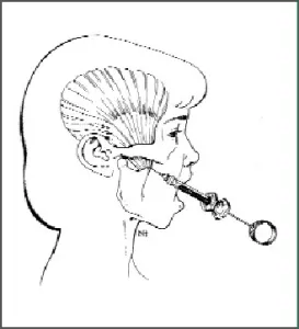
One way to determine if the pain is caused by the temporal tendon is to put your finger in your mouth and gently push outward on the cheek, at the top of the jaw.

Symptoms include:
- Constant aching behind the eye
- Sensitivity to bright light (photophobia)
- Intense headache, lateral temple headaches
- Upper and lower molar teeth pain
- Cheeks swell up
- Restricted jaw movement AKA trismus
- Ear pain and pressure
- Radiation of pain from the cheek to eye
- Sometimes; Nausea, vomiting, visual disturbances
What are the causes & diagnosis of Temporalis Tendonitis?
Temporalis Tendonitis is often associated with prolonged mouth opening (such as visits to the dentist), increased
stress, tooth grinding, direct trauma to the Temporalis muscle, excessive gum chewing. In rare cases a condition called Coronoid Process Hyperplasia may be the cause of Temporal Tendinitis. The diagnosis of Temporal Tendinitis by palpation of the tendon as it inserts into the coronoid process. Injection of local anesthetic into this region should alleviate the pain and this is regarded as the definitive diagnostic test.
How is Temporal Tendinitis Treated?
To treat Temporal Tendinitis effectively, both dental and medical knowledge in the areas of teeth, soft tissue, and TM joint function is required. If the patient has jaw joint pain and soft tissue pain, the clinician must determine which is primary and which is secondary in the diagnostic stream. In particular, it is crucial to first assess the condition of the jaw joint and its muscles and tendons before proceeding to equilibrate the teeth.
Once Temporal Tendinitis is diagnosed properly, we may recommend treatment with injections of a mixture of anesthetics and pain-blockers (Anti-inflammatories). This procedure is typically repeated for a series of three or more treatments.

What is Coronoid Process Hyperplasia (CPH)?
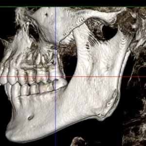
Coronoid Process Hyperplasia (CPH) is a rare condition that causes mouth opening limitations, otherwise known as trismus. The picture on the right shows the coronoid process in red. The elongated coronoid processes impinge on the medial side of the zygomatic arch (cheek bone) when opening the mouth and moving the jaw side to side. The limited range of motion of the mandible leads to pain and limited mouth opening (trismus).
How do you know if you have Coronoid Process Hyperplasia (CPH) as the primary cause of your pain or limited mouth opening?
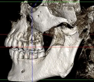
Patients with trismus due to CPH do not have any definitive symptoms such as temporomandibular joint (TMJ pain) or clicking or popping. Therefore the diagnosis of Coronoid process hyperplasia is very difficult. CT scans can help with diagnosis. As you can see in our patient’s CT scan on the right, the coronoid processes are very large in this patient. He had difficulty in mouth opening and pain and swelling in the temples.
How is Coronoid Process Hyperplasia Treated?
Coronoid Process Hyperplasia is treated with a procedure called coronoidectomy. In this procedure a small incision is made inside the oral cavity and the Coronoid Process is exposed. This elongated bone is then shaved down to a normal height (Scan on the above left). The muscle then will reattach to the new position of the coronoid process. Physical therapy after the surgery is important part of healing to ensure adequate mouth opening.